Papers
The discoveries that advance science
The exceptional discoveries in oncology and cancer research made by scientists and clinicians in Beijing are captured by a suite of Cell Press journals, among which we highlight several representatives showcasing the diverse topics and broad scope of these achievements.

Tumor-Repopulating Cells Induce PD-1 Expression in CD8+ T Cells by Transferring Kynurenine and AhR Activation
Cancer Cell
VOLUME 33, ISSUE 3, P480-494.E7, 03/12/2018,
Read the paper on Cell.comBo Huang
- Department of Immunology & National Key Laboratory of Medical Molecular Biology, Institute of Basic Medical Sciences, Chinese Academy of Medical Sciences (CAMS) & Peking Union Medical College, Beijing, China
- Clinical Immunology Center, CAMS, Beijing, China
- Department of Biochemistry & Molecular Biology, Tongji Medical College, Huazhong University of Science & Technology, Wuhan, China
Abstract
Despite the clinical successes fostered by immune checkpoint inhibitors, mechanisms underlying PD-1 upregulation in tumor-infiltrating T cells remain an enigma. Here, we show that tumor-repopulating cells (TRCs) drive PD-1 upregulation in CD8+ T cells through a transcellular kynurenine (Kyn)-aryl hydrocarbon receptor (AhR) pathway. Interferon-γ produced by CD8+ T cells stimulates release of high levels of Kyn produced by TRCs, which is transferred into adjacent CD8+ T cells via the transporters SLC7A8 and PAT4. Kyn induces and activates AhR and thereby upregulates PD-1 expression. This Kyn-AhR pathway is confirmed in both tumor-bearing mice and cancer patients and its blockade enhances antitumor adoptive T cell therapy efficacy. Thus, we uncovered a mechanism of PD-1 upregulation with potential tumor immunotherapeutic applications.
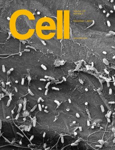
The Mevalonate Pathway Is a Druggable Target for Vaccine Adjuvant Discovery
Cell
VOLUME 175, ISSUE 4, P1059-1073.E21, 11/01/2018,
Read the paper on Cell.comWanli Liu
MOE Key Laboratory of Protein Sciences, School of Life Sciences, Tsinghua University, Beijing, China Institute for Immunology and School of Medicine, Tsinghua University, Beijing, China
Yan Shi
Institute for Immunology and School of Medicine, Tsinghua University, Beijing, China Institute Department of Microbiology, Immunology and Infectious Diseases, Snyder Institute, University of Calgary, Calgary, AB, Canada
Yonghui Zhang
School of Pharmaceutical Sciences, MOE Key Laboratory of Bioorganic Phosphorus Chemistry and Chemical Biology, Tsinghua University, Beijing, China
Joint Graduate Program of Peking-Tsinghua-NIBS, School of Life Sciences, Tsinghua University, Beijing, China
Collaborative Innovation Center for Biotherapy, Sichuan University, Chengdu, Sichuan, China
Abstract
Motivated by the clinical observation that interruption of the mevalonate pathway stimulates immune responses, we hypothesized that this pathway may function as a druggable target for vaccine adjuvant discovery. We found that lipophilic statin drugs and rationally designed bisphosphonates that target three distinct enzymes in the mevalonate pathway have potent adjuvant activities in mice and cynomolgus monkeys. These inhibitors function independently of conventional “danger sensing.” Instead, they inhibit the geranylgeranylation of small GTPases, including Rab5 in antigen-presenting cells, resulting in arrested endosomal maturation, prolonged antigen retention, enhanced antigen presentation, and T cell activation. Additionally, inhibiting the mevalonate pathway enhances antigen-specific anti-tumor immunity, inducing both Th1 and cytolytic T cell responses. As demonstrated in multiple mouse cancer models, the mevalonate pathway inhibitors are robust for cancer vaccinations and synergize with anti-PD-1 antibodies. Our research thus defines the mevalonate pathway as a druggable target for vaccine adjuvants and cancer immunotherapies.
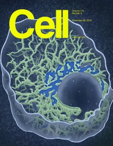
Mutational Landscape of Secondary Glioblastoma Guides MET-Targeted Trial in Brain Tumor Cell
Cell
VOLUME 175, ISSUE 6, P1665-1678.E18, 11/29/2018,
Read the paper on Cell.comXiaolong Fan
Laboratory of Neuroscience and Brain Development, Beijing Key Laboratory of Gene Resource and Molecular Development, Beijing Normal University, Beijing, China
Jiguang Wang
Department of Chemical and Biological Engineering, The Hong Kong University of Science and Technology, Hong Kong, China
Division of Life Science, The Hong Kong University of Science and Technology, Hong Kong, China
Center of Systems Biology and Human Health, The Hong Kong University of Science and Technology, Hong Kong, China
Tao Jiang
Beijing Neurosurgical Institute, Capital Medical University, Beijing, China
Department of Neurosurgery, Beijing Tiantan Hospital, Capital Medical University, Beijing, China
Center of Brain Tumor, Beijing Institute for Brain Disorders, Beijing, China
China National Clinical Research Center for Neurological Diseases, Beijing, China
Abstract
Low-grade gliomas almost invariably progress into secondary glioblastoma (sGBM) with limited therapeutic option and poorly understood mechanism. By studying the mutational landscape of 188 sGBMs, we find significant enrichment of TP53 mutations, somatic hypermutation, MET-exon-14-skipping (METex14), PTPRZ1-MET (ZM) fusions, and MET amplification. Strikingly, METex14 frequently co-occurs with ZM fusion and is present in ∼14% of cases with significantly worse prognosis. Subsequent studies show that METex14 promotes glioma progression by prolonging MET activity. Furthermore, we describe a MET kinase inhibitor, PLB-1001, that demonstrates remarkable potency in selectively inhibiting MET-altered tumor cells in preclinical models. Importantly, this compound also shows blood-brain barrier permeability and is subsequently applied in a phase I clinical trial that enrolls MET-altered chemo-resistant glioma patients. Encouragingly, PLB-1001 achieves partial response in at least two advanced sGBM patients with rarely significant side effects, underscoring the clinical potential for precisely treating gliomas using this therapy.
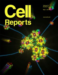
Dissecting the Single-Cell Transcriptome Network Underlying Gastric Premalignant Lesions and Early Gastric Cancer
Cell Reports
VOLUME 27, ISSUE 6, P1934-1947.E5, 05/07/2019,
Read the paper on Cell.comShao Li
MOE Key Laboratory of Bioinformatics, TCM-X Centre/Bioinformatics Division, BNRIST/Department of Automation, Tsinghua University, Beijing, China
Abstract
Intestinal-type gastric cancer is preceded by premalignant lesions, including chronic atrophic gastritis and intestinal metaplasia. In this study, we constructed a single-cell atlas for 32,332 high-quality cells from gastric antral mucosa biopsies of patients spanning a cascade of gastric premalignant lesions and early gastric cancer (EGC) using single-cell RNA sequencing. We then constructed a single-cell network underlying cellular and molecular characteristics of gastric epithelial cells across different lesions. We found that gland mucous cells tended to acquire an intestinal-like stem cell phenotype during metaplasia, and we identified OR51E1 as a marker for unique endocrine cells in the early-malignant lesion. We also found that HES6 might mark the pre-goblet cell cluster, potentially aiding identification of metaplasia at the early stage. Finally, we identified a panel of EGC-specific signatures, with clinical implications for the precise diagnosis of EGC. Our study offers unparalleled insights into the human gastric cellulose in premalignant and early-malignant lesions.

Genomic and Transcriptomic Profiling of Combined Hepatocellular and Intrahepatic Cholangiocarcinoma Reveals Distinct Molecular Subtypes
Cancer Cell
VOLUME 35, ISSUE 6, P932-947.E8, 06/10/2019,
Read the paper on Cell.comHidewaki Nakagawa
Laboratory for Cancer Genomics, RIKEN Center for Integrative Medical Sciences, Yokohama, Japan
Mu-Sheng Zeng
State Key Laboratory of Oncology in South China, Collaborative Innovation Center for Cancer Medicine, Sun Yat-Sen University Cancer Center, Guangzhou, China
Fan Bai
Biomedical Pioneering Innovation Center (BIOPIC) and Translational Cancer Research Center, School of Life Sciences, First Hospital, Peking University, Beijing, China
Ning Zhang
Biomedical Pioneering Innovation Center (BIOPIC) and Translational Cancer Research Center, School of Life Sciences, First Hospital, Peking University, Beijing, China
Laboratory of Cancer Cell Biology, Tianjin Medical University Cancer Institute and Hospital, Tianjin, China
Abstract
We performed genomic and transcriptomic sequencing of 133 combined hepatocellular and intrahepatic cholangiocarcinoma (cHCC-ICC) cases, including separate, combined, and mixed subtypes. Integrative comparison of cHCC-ICC with hepatocellular carcinoma and intrahepatic cholangiocarcinoma revealed that combined and mixed type cHCC-ICCs are distinct subtypes with different clinical and molecular features. Integrating laser microdissection, cancer cell fraction analysis, and single nucleus sequencing, we revealed both mono- and multiclonal origins in the separate type cHCC-ICCs, whereas combined and mixed type cHCC-ICCs were all monoclonal origin. Notably, cHCC-ICCs showed significantly higher expression of Nestin, suggesting Nestin may serve as a biomarker for diagnosing cHCC-ICC. Our results provide important biological and clinical insights into cHCC-ICC.
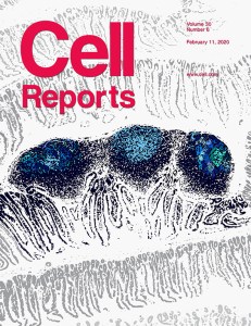
Secreted Pyruvate Kinase M2 Promotes Lung Cancer Metastasis through Activating the Integrin Beta1/FAK Signaling Pathway
Cell Reports
VOLUME 30, ISSUE 6, P1780-1797.E6, 02/11/2020,
Read the paper on Cell.comYongzhang Luo
Cancer Biology Laboratory, School of Life Sciences, Tsinghua University, Beijing, China
The National Engineering Laboratory for Anti-Tumor Protein Therapeutics, Tsinghua University, Beijing, China
Beijing Key Laboratory for Protein Therapeutics, Tsinghua University, Beijing, China
Abstract
Cancer cell-derived secretomes have been documented to play critical roles in cancer progression. Intriguingly, alternative extracellular roles of intracellular proteins are involved in various steps of tumor progression, which can offer strategies to fight cancer. Herein, we identify lung cancer progression-associated secretome signatures using mass spectrometry analysis. Among them, PKM2 is verified to be highly expressed and secreted in lung cancer cells and clinical samples. Functional analyses demonstrates that secreted PKM2 facilitates tumor metastasis. Furthermore, mass spectrometry analysis and functional validation identify integrin β1 as a receptor of secreted PKM2. Mechanistically, secreted PKM2 directly bound to integrin β1 and subsequently activated the FAK/SRC/ERK axis to promote tumor metastasis. Collectively, our findings suggest that PKM2 is a potential serum biomarker for diagnosing lung cancer and that targeting the secreted PKM2-integrin β1 axis can inhibit lung cancer development, which provides evidence of a potential therapeutic strategy in lung cancer.

Single-Cell Analyses Inform Mechanisms of Myeloid-Targeted Therapies in Colon Cancer
Cell
VOLUME 181, ISSUE 2, P442-459.E29, 04/16/2020,
Read the paper on Cell.comZhanlong Shen
Department of Gastroenterological Surgery, Peking University People’s Hospital, Beijing, China
Jackson G. Egen
Department of Inflammation and Oncology and Genome Analysis Unit, Amgen Research, Amgen Inc., South San Francisco, CA, USA
Zemin Zhang
Beijing Advanced Innovation Center for Genomics, Peking-Tsinghua Center for Life Sciences, Peking University, Beijing, China
BIOPIC and School of Life Sciences, Peking University, Beijing, China
Xin Yu
Department of Inflammation and Oncology and Genome Analysis Unit, Amgen Research, Amgen Inc., South San Francisco, CA, USA
Abstract
Single-cell RNA sequencing (scRNA-seq) is a powerful tool for defining cellular diversity in tumors, but its application toward dissecting mechanisms underlying immune-modulating therapies is scarce. We performed scRNA-seq analyses on immune and stromal populations from colorectal cancer patients, identifying specific macrophage and conventional dendritic cell (cDC) subsets as key mediators of cellular cross-talk in the tumor microenvironment. Defining comparable myeloid populations in mouse tumors enabled characterization of their response to myeloid-targeted immunotherapy. Treatment with anti-CSF1R preferentially depleted macrophages with an inflammatory signature but spared macrophage populations that in mouse and human expresses pro-angiogenic/tumorigenic genes. Treatment with a CD40 agonist antibody preferentially activated a cDC population and increased Bhlhe40 + Th1-like cells and CD8 + memory T cells. Our comprehensive analysis of key myeloid subsets in human and mouse identifies critical cellular interactions regulating tumor immunity and defines mechanisms underlying myeloid-targeted immunotherapies currently undergoing clinical testing.

Integrative Proteomic Characterization of Human Lung Adenocarcinoma
Cell
VOLUME 182, ISSUE 1, P245-261.E17, 07/09/2020,
Read the paper on Cell.comYi Wang
State Key Laboratory of Proteomics, Beijing Proteome Research Center, National Center for Protein Sciences (Beijing), Beijing Institute of Lifeomics, Beijing, China
Jing Li
Department of Bioinformatics and Biostatistics, School of Life Sciences and Biotechnology, Shanghai Jiao Tong University, Shanghai, China
Fuchu He
State Key Laboratory of Proteomics, Beijing Proteome Research Center, National Center for Protein Sciences (Beijing), Beijing Institute of Lifeomics, Beijing, China
Ting Xiao
State Key Laboratory of Molecular Oncology, Department of Etiology and Carcinogenesis, National Cancer Center/National Clinical Research Center for Cancer/Cancer Hospital, Chinese Academy of Medical Sciences and Peking Union Medical College, Beijing, China
Minjia Tan
State Key Laboratory of Drug Research, Shanghai Institute of Materia Medica, Chinese Academy of Sciences, Shanghai, China
University of the Chinese Academy of Sciences, Beijing, China
Abstract
Genomic studies of lung adenocarcinoma (LUAD) have advanced our understanding of the disease’s biology and accelerated targeted therapy. However, the proteomic characteristics of LUAD remain poorly understood. We carried out a comprehensive proteomics analysis of 103 cases of LUAD in Chinese patients. Integrative analysis of proteome, phosphoproteome, transcriptome, and whole-exome sequencing data revealed cancer-associated characteristics, such as tumor-associated protein variants, distinct proteomics features, and clinical outcomes in patients at an early stage or with EGFR and TP53 mutations. Proteome-based stratification of LUAD revealed three subtypes (S-I, S-II, and S-III) related to different clinical and molecular features. Further, we nominated potential drug targets and validated the plasma protein level of HSP 90β as a potential prognostic biomarker for LUAD in an independent cohort. Our integrative proteomics analysis enables a more comprehensive understanding of the molecular landscape of LUAD and offers an opportunity for more precise diagnosis and treatment.
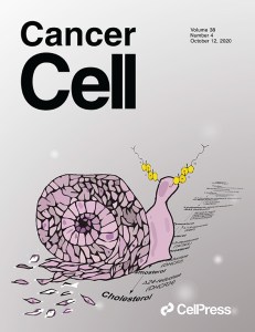
Hematopoietic Progenitor Kinase1 (HPK1) Mediates T Cell Dysfunction and Is a Druggable Target for T Cell-Based Immunotherapies
Cancer Cell
VOLUME 38, ISSUE 4, P551-566.E11, 10/12/2020,
Read the paper on Cell.comLai Wei
State Key Laboratory of Ophthalmology, Zhongshan Ophthalmic Center, Sun Yat-sen University, Guangzhou, China
Xuebin Liao
School of Pharmaceutical Sciences, Key Laboratory of Bioorganic Phosphorus Chemistry and Chemical Biology (Ministry of Education), Beijing Advanced Innovation Center for Human Brain Protection, Tsinghua University, Beijing, China
Abstract
Ameliorating T cell exhaustion and enhancing effector function are promising strategies for the improvement of immunotherapies. Here, we show that the HPK1-NFκB-Blimp1 axis mediates T cell dysfunction. High expression of MAP4K1 (which encodes HPK1) correlates with increased T cell exhaustion and with worse patient survival in several cancer types. In MAP4K1 KO mice, tumors grow slower than in wild-type mice and infiltrating T cells are less exhausted and more active and proliferative. We further show that genetic depletion, pharmacological inhibition, or proteolysis targeting chimera (PROTAC)-mediated degradation of HPK1 improves the efficacy of CAR-T cell-based immunotherapies in diverse preclinical mouse models of hematological and solid tumors. These strategies are more effective than genetically depleting PD-1 in CAR-T cells. Thus, we demonstrate that HPK1 is a mediator of T cell dysfunction and an attractive druggable target to improve immune therapy responses.

PPARα Inhibition Overcomes Tumor-Derived Exosomal Lipid-Induced Dendritic Cell Dysfunction
Cell Reports
VOLUME 33, ISSUE 3, 108278, 10/20/2020,
Read the paper on Cell.comWenfeng Zeng
Protein and Peptide Pharmaceutical Laboratory, Institute of Biophysics, Chinese Academy of Sciences, Beijing, China
University of Chinese Academy of Sciences, Beijing, China
Lingtao Jin
Department of Anatomy and Cell Biology, College of Medicine, University of Florida, Gainesville, FL, USA
Wei Liang
Protein and Peptide Pharmaceutical Laboratory, Institute of Biophysics, Chinese Academy of Sciences, Beijing, China
University of Chinese Academy of Sciences, Beijing, China
Abstract
Dendritic cells (DCs) orchestrate the initiation, programming, and regulation of anti-tumor immune responses. Emerging evidence indicates that the tumor microenvironment (TME) induces immune dysfunctional tumor infiltrating DCs (TIDCs), characterized with both increased intracellular lipid content and mitochondrial respiration. The underlying mechanism, however, remains largely unclear. Here, we report that fatty acid-carrying tumor-derived exosomes (TDEs) induce immune dysfunctional DCs to promote immune evasion. Mechanistically, peroxisome proliferator activated receptor (PPAR) α responds to the fatty acids delivered by TDEs, resulting in excess lipid droplet biogenesis and enhanced fatty acid oxidation (FAO), culminating in a metabolic shift toward mitochondrial oxidative phosphorylation, which drives DC immune dysfunction. Genetic depletion or pharmacologic inhibition of PPARα effectively attenuates TDE-induced DC-based immune dysfunction and enhances the efficacy of immunotherapy. This work uncovers a role for TDE-mediated immune modulation in DCs and reveals that PPARα lies at the center of metabolic-immune regulation of DCs, suggesting a potential immunotherapeutic target.

